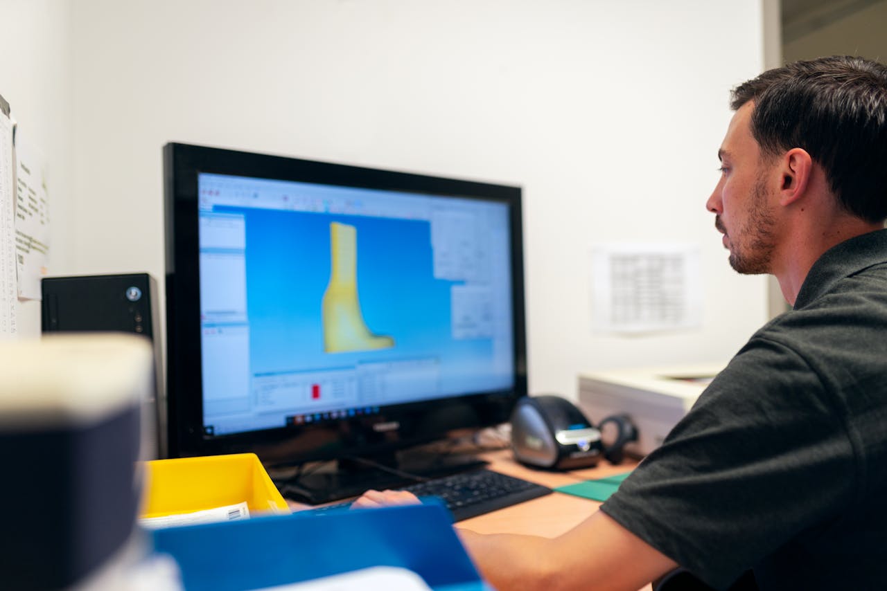
Could 3D mapping identify misalignments before pain begins? With this tech, you receive a crisp scan of bone and joint placement, so tiny shifts pop up sooner. You can actually see any imbalances in posture or movement immediately on the screen. Several clinics use 3D scans to monitor small shifts that often escape a normal exam. Early checks allow you to begin simple fixes, like posture aids or stretches, before pain emerges. This technique applies to multiple areas of the body, from your back to your knees. If you want to maintain an active lifestyle and avoid debilitating joint or muscle problems in the future, frequent 3D scans can be a smart way to identify these issues before they become severe. Read on to see how it works.
Key Takeaways
- Before you even notice pain, 3D mapping can detect misalignments so you can be proactive and prevent long-term issues.
- It offers precision scans of your body or feet, assisting practitioners in developing customized treatment or orthotic plans specific to your anatomy.
- Using 3D mapping to detect asymmetries and muscular imbalances early on promotes overall health, comfort, and performance.
- 3D mapping is a more holistic and accessible solution than traditional imaging. It provides instant feedback and faster results.
- By doing regular 3D mapping, you and your healthcare provider can monitor your progress, stay ahead of any misalignments before the pain starts, and keep you in peak condition for years to come.
- By incorporating 3D mapping into your healthcare or training routine, you can minimize your risk of injury and promote sustainable health with data-driven personalized care.

What is 3D Mapping?
3D mapping is a virtual mapping technology that captures objects, bodies, or spaces in detailed 3D models. In healthcare, you might encounter this tech employed for 3D body scanning to record the complete form and anatomy of your feet, spine, or joints. These mapped models are not just pictures; they are physically accurate replicas constructed with spatial information, so they depict your actual form and surface. This technique is crucial for posture analysis and alignment checks because it provides detailed data that is difficult to achieve with traditional instruments.
The Technology
3D mapping utilizes advanced imaging technologies like laser scanning, structured light, and photogrammetry. They scan and capture the minutest variations of your body’s surface to millimeter precision. This degree of accuracy implies that even mild misalignments can be detected before they develop into discomfort.
Specialized software then uses this information to construct a digital model. The software can analyze, quantify, and compare your anatomy like no 2D images ever could. Most of these are safe systems, using either non-invasive light or low-energy scanners, so no radiation is involved.
The Process
- You stand or sit in a fixed position, depending on whether it is being scanned.
- The scanner, either mounted on a frame or held by a specialist, sweeps over the space.
- The system captures thousands of data points in mere seconds.
- Calibration occurs before each scan to ensure the device reads accurate measurements.
- Software constructs the 3D model and verifies errors.
If you sway or jiggle during the 3D body scan, the results could be fuzzy or inaccurate, so maintaining proper posture and immobility is crucial. Calibration is key to achieving precise 3D imaging. Without it, your model may not appear life-like. In contrast to traditional approaches that might require multiple hours or visits, advanced 3D imaging is rapid, with each scan taking under five minutes, saving you both time and headache.
The Difference
3D Mapping | X-rays | MRIs | |
View | 3D, full surface | 2D, limited | 2D/3D, deep |
Radiation | None/Low | Yes | None |
Detail | High (surface) | Bone-focused | Soft tissue |
Time | Fast (minutes) | Moderate | Slow |
Different from X-rays or MRIs, 3D body scans display the entire outer shape, providing a clearer glimpse of how your posture alignment is working. The system can provide immediate feedback, highlighting problems while you’re still in the office. This simplifies the conversation between you and your provider about what’s occurring. For most people, the scan is easy and unstrained—no claustrophobic tubes or twisted limbs.
How 3D Mapping Detects Misalignments
3D mapping detects misalignments prior to symptoms beginning by constructing a comprehensive map of your body’s alignment, posture, and movement. With advanced 3D imaging, digital twins, and detailed biomechanical analysis, this tech catches the little shifts that can cause pain or chronic issues down the road. Early detection through posture analysis provides you with a greater chance of preventing pain in its tracks, which translates to healthier living over time.
1. Capturing Surface Data
3D foot scans collect surface data by scanning the complete contour of your feet as you stand or are in motion. This detects even minor changes in the height or angle of the foot, which can indicate early misalignments. The scan’s precision typically comes from digital imaging, which employs thousands of data points to create an accurate digital map.
This accurate surface data matters because it reveals places where your foot or body drifts away from normal alignment, even when you feel fine. It’s crucial for creating custom orthotics that mold perfectly to your foot, not just a “generic” form. That way, your shoes or supports can align with your actual needs.
2. Creating a Digital Twin
A digital twin is a virtual representation of your body, constructed from 3D scans. This model is specific to you and details your actual posture and alignment. It allows doctors to detect minor misalignments in your spine or feet that can be overlooked by the naked eye.
With a digital twin, you receive care tailored to your body, not just averages. This model could be updated over time to monitor if your alignment shifts, allowing you to make smarter treatment decisions as you go.
3. Identifying Asymmetries
3D mapping detects misalignments in your feet or spine, such as one side being higher or more rotated than the other. These minor misalignments can knock your entire body out of whack and create joint or muscle strain. Even if you’re not feeling discomfort yet, these concealed imbalances can cause aches or issues down the line.
To this moment, we’ve been focused on how 3D mapping actually detects misalignments. For instance, if the 3D mapping reveals your left foot rolls in more than your right, custom orthotics can be created to correct that discrepancy. This step can prevent small things from becoming bigger health concerns.
4. Analyzing Biomechanics
3D mapping allows practitioners to analyze your motion, the way your joints bend, your spine aligns, and weight shifts during your step. This deep dive into biomechanics ties in directly to joint health. If your motion is misaligned, joints can wear prematurely and cause discomfort.
About: How 3D Mapping Reveals Misalignments and it’s key for injury prevention too, because knowing your patterns of movement will identify weak spots before they become painful or damaged.
5. Quantifying Deviations
3D mapping isn’t just for pictures. It provides us with quantitative data that reveals the degree to which your alignment diverges from ideal. This information is valuable for monitoring your improvements and ensuring interventions, such as orthotics or exercise, are effective.
Accurate figures help you detect actual progress, not just wishful thinking. It’s this precision that makes 3D mapping so helpful for long-term health.
Beyond Bones: A Holistic View
3D body scanning allows us to see so much more than just bones. To catch postural imbalances before you ache, you have to see the whole system: muscles, joints, posture, and movement. This isn’t just about the skeleton; it’s the tissues, tendons, cartilage, and ligaments going in harmony. A holistic perspective provides a richer, more practical understanding.
- Muscle imbalance often hides in early joint changes.
- Ignoring muscle health can lead to under- or late misalignment identification.
- Soft tissues affect both comfort and movement.
- Caring for your muscles and tendons early can save you from long-term pain.
Muscular Imbalance
Muscular imbalance occurs when the muscles on either side of a joint are disproportionately stronger or tighter, which can lead to joint strain or abuse. This misalignment can result in back discomfort and other issues over time, even without an injury. Utilizing advanced 3D imaging techniques, such as 3D body scans, allows for a detailed view of where muscles are not functioning properly, rather than just identifying misaligned bones.
3D mapping provides vivid, layered images that reveal weak or tight spots, while decalcification and deep tissue imaging capture soft tissue specifics for a more complete understanding of the musculoskeletal system. Neglecting muscular imbalances can lead to overlooking the source of pain syndromes, as most pain issues trace their roots back to minor, early muscle movements that standard imaging may miss.
Addressing muscle balance early through posture alignment therapy can inhibit or prevent pain from taking hold, ensuring proper alignment and enhancing overall health. By leveraging 3D body scanning technology, you can gain insights into your posture and work towards achieving balance and symptom relief.
Postural Patterns
3D mapping can detect patterns in your posture and movement. If you slouch or tilt your pelvis, it is likely to stress your joints even if you don’t notice it now. Research reveals that pelvic tilt and sagittal imbalance are significant factors in posture problems and pain.
Bad posture can grind down joints or even cause them to change shape. With three-dimensional image stacks, you can observe these changes in detail, aiding early interventions.
A good postural check not only reveals what’s off, it helps you fix it. Keeping you to maintain good posture makes these changes stick.
Gait Analysis
3D mapping allows you to observe the motion of each segment of your body during locomotion. Even subtle gait changes can reveal misalignments you don’t detect. These subtle changes can strain joints, muscles, or tendons in the long run.
If you can catch gait issues early, you can use orthotics or change your routine before the pain begins. 3D gait scans are crucial in fine-tuning orthotic design, ensuring your support matches your specific stride.
Monitoring your stride longitudinally assists you in identifying and correcting issues, maintaining your mobility, and making you healthier.

From Data to Prevention
3D body scanning provides a transparent view into your body’s alignment prior to the onset of pain. This advanced 3D imaging technology monitors minor postural shifts, such as vertebral rotation beyond 10 degrees or pelvic tilt in excess of 4 millimeters, which are associated with increased pain and discomfort. With this data, you don’t have to wait for symptoms to appear. You can act early, reduce your risk, and stay moving strong.
- Spot abnormal posture early, like sagittal imbalance > 5 mm
- Create personalized workouts or therapeutic regimens derived from your specific data.
- Adjust orthotics or supports quickly if changes are found
- Track small shifts over time to prevent larger issues
- Turn your data into prevention.
Personalized Plans
3D body scanning allows you to have a care plan designed specifically for you, not a generic plan. When your posture scans detect a change, like a sagittal imbalance of over 6 mm, it informs your provider in designing your individualized treatment plan. This could include custom orthotics, exercises, or posture alignment therapy. Custom orthotics molded to your mapping data translate to improved support and comfort. They assist in distributing pressure equally, minimizing pain, and increasing your mobility. Over time, these 3D scans help your team adjust your plan as your body shifts, which keeps your alignment in check and prevents issues before they arise.
Proactive Adjustments
With advanced 3D imaging, your orthotic tweaks can keep up with your body’s changes. If your 3D body scan reveals a new tilt or shift in your neck alignment, you can adjust your supports immediately instead of waiting for pain to develop. This timely intervention prevents minor problems from becoming major ones. Routine posture analysis examinations allow you and your clinician to identify non-linear shifts or anomalous trends, such as those exhibited in fibromyalgia, before they escalate into larger issues. Your voice counts as well; when you share what you feel and notice, you help shape your own care for better results.
Performance Enhancement
- Add mapping checks to your training schedule for early warning signs.
- Take advantage of your custom alignment data to fine-tune form and posture.
- Pick orthotics designed for your specific needs to reduce the risk of injury.
- Monitor progress and modify your schedule as your body acclimates.
- Share mapping results with your coach or therapist for better feedback.
Alignment is not just important for pain relief; it plays a crucial role in performance and longevity. Good posture, shaped by precise 3D body scans, can amplify your power, agility, and pace. Athletes utilizing posture analysis and custom orthotics tend to experience fewer injuries and more consistent improvement.
The Evidence for Early Detection
Our Evidence for Early Detection 3D mapping technology is transforming the way you and your care team identify postural imbalances before the onset of pain. The science supports this. Digital and 3D body scanning now assist with early detection, improved monitoring, and better communication between you and your physician. New science reveals that 3D pain mapping provides you and your care team with more objective ways to monitor pain longitudinally, even remotely, using advanced 3D imaging tools.
Clinical Support
It turns into compelling clinical evidence for the use of 3D mapping in identifying misalignments. Digital pain mapping and 3D charts enable you to log pain as it occurs or over days or even months, making it far easier to identify trends that could transform into chronic issues. When doctors and tech experts collaborate, they bring both clinical wisdom and cutting-edge technology to your care, so you receive a more comprehensive snapshot of what’s going on in your body. This collaborative effort allows 3D mapping to identify subtle changes that could be overlooked with previous approaches.
3D mapping provides your care team with enhanced clarity during diagnosis. It assists them in identifying precisely where imbalances begin and how they may propagate or transform over time. Evidence-based use of 3D mapping means your treatment plan is based on real data, not guesswork, which helps your care stay focused and precise.
Study Reference | Findings | Clinical Impact |
Test-Retest Reliability (2022) | 3D pain mapping improved location reliability | Better early detection and monitoring |
| Sagittal Imbalance Study (2021) | Greater than 5 millimeters of imbalance equals a higher pain risk | About | The Case for Early Intervention
EMA Feasibility Review, 2020. EMAs monitor pain on a daily basis remotely. The potential for early intervention.
Predictive Power
3D mapping provides your care team with an opportunity to anticipate potential misalignments before you experience discomfort. It leverages X-rays and digital monitoring to detect minor changes in posture, such as a tilt or imbalance, that might otherwise develop into something more serious. For instance, studies revealed that a pelvic tilt in excess of 3 mm can dramatically spike pain scores down the road.
With this foresight, your doctor can take preventive action early, rather than waiting for you to start hurting. Data-driven insights enable you and your care team to make decisions around lifestyle changes or treatments that can keep pain at bay. Today, predictive analytics from such images is crucial in preventing chronic pain, particularly among high-risk populations such as women with fibromyalgia, who experience higher rates of chronic pain.
Known Limitations
As for the evidence in favor of early detection, 3D mapping isn’t perfect. It can’t prove everything, of course, particularly in cases of complex pain or psychological overlay. Occasionally, a clear scan isn’t sufficient. Your physician might have to incorporate additional tests or use pain drawings to obtain the complete picture.
Continuous research is on the way to make 3D mapping even more trustworthy. Managing expectations is important. 3D mapping can help steer early treatment, but it’s not a miracle cure for all cases.
The Future of Predictive Health
3D mapping is carving out a fresh route for health care by providing you with foresight into your body before pain sets in. It can perform a 3D body scan of your spine, shoulders, or other joints in high resolution, revealing tiny shifts or twists that you can’t sense yet. These scans, when coupled with data from wearable trackers or your medical records, assist in constructing a more comprehensive picture of your health. This gives you more opportunity to address minor issues before they become major.
AI is accelerating the speed and accuracy with which this data is interpreted. AI can detect patterns in your posture scans that are simple to overlook if you do it yourself. For instance, machine learning can read changes in your cervical alignment and indicate whether you are at risk of future neck pain. AI utilizes models that fragment your body, such as finite element models, to observe how each fragment moves and responds to stress. This helps identify what may go awry and why. With R-squared values between 0.298 and 0.549, these models already provide a moderate to strong association between what is observed in your scans and what you report about pain or function.
Integrating 3D body scanning into a broader health plan is crucial. Your shape is only one piece of the picture. When you throw in mental and social data—stress, sleep, and your daily habits—your care becomes more holistic. Predictive models now use information from many sources: health records, fitness trackers, and even self-reports to figure out which treatments work best for you. This is useful for selecting who may benefit most from specific treatments, such as cervical lordosis rehabilitation, and for monitoring whether you’re hitting important progress markers in your pain or mobility.
For you, this means care can be more personal, with less guesswork. Doctors can leverage these tools not just to catch misalignments early, but to select treatments with a greater likelihood of success for your individual case. As these models expand, higher-level scans such as MRI and CT will be incorporated, rendering predictions more precise and care more personalized. Down this road lie less pain, quicker healing, and more affordable health care for all.
Conclusion
3D mapping lets you see your body before pain arrives. It identifies tiny misalignments and highlights where things could soon derail. This means you can spot bone and joint changes early. You receive data that helps you collaborate with your care team, not merely respond to pain. They are leveraging this tech to better plan workouts, correct motion, and protect joints. Early checks with 3D mapping can help you keep your body strong for the long run. More control and less guesswork. Try a scan, talk with your provider, and see what it uncovers. That next step might be the beginning of change.
Frequently Asked Questions
Can 3D mapping detect misalignments before I feel pain?
Yes. They utilize 3D body scanning to map how your body is misaligned before symptoms or pain even begin, allowing you to take action before it is too late.
How does 3D mapping work for early detection?
3D body scanning analyzes precise images of your body’s structure, identifying subtle misalignments and changes in posture alignment that are hard to detect with conventional techniques.
Is 3D mapping only for bones?
No. 3D body scanning is holistic, incorporating muscles, joints, and soft tissues for a complete profile of your posture analysis and balance.
Why is early detection of misalignments important?
Early detection through posture analysis and 3D body scanning helps you fix issues before they become painful or injurious.
Can 3D mapping help prevent injuries?
Yes. Because it can identify postural imbalances before pain kicks in, 3D body scanning lets you take personalized preventative actions and minimizes your chances of a sports injury or chronic ache.
Is 3D mapping safe?
Yes. 3D body scanning is safe for individuals of all ages and health conditions, as it employs non-invasive imaging methods.
Who should consider 3D mapping for alignment checks?
Anyone who wants to be proactively healthy, particularly athletes or those with repetitive stress, can benefit from posture analysis. It’s excellent for tracking neck posture and addressing potential postural imbalances early.
Struggling With Foot, Back, or Knee Pain? Find Answers With 3D Foot Mapping at The Shoe Doctor.
If you deal with constant back or knee pain, your feet could be the real source of the problem. Even small misalignments in your foot structure can throw off your entire body’s balance, putting strain on your joints and muscles. At The Shoe Doctor, we use advanced 3D foot mapping to pinpoint these imbalances with incredible precision.
Our detailed foot-mapping process captures how you stand, move, and distribute pressure, allowing us to create truly custom orthotics that correct alignment, relieve pressure points, and restore natural movement. With more than 20 years of experience, Russell combines cutting-edge technology with expert craftsmanship to design orthotics that do more than add comfort—they help prevent pain from returning.
Through our partnership with the Spine & Injury Medical Center in San Jose, we take a full-body approach to relief, ensuring your posture, gait, and foot mechanics all work together for lasting results.
If you’re in the South Bay Area, book your free consultation today. Let The Shoe Doctor use 3D foot mapping to help you move comfortably and confidently again.
Disclaimer
The materials available on this website are for informational and entertainment purposes only and are not intended to provide medical advice. You should contact your doctor for advice concerning any particular issue or problem. You should not act or refrain from acting based on any content included in this site without seeking medical or other professional advice. The information presented on this website may not reflect the most current medical developments. No action should be taken in reliance on the information contained on this website, and we disclaim all liability for actions taken or not taken based on any or all of the contents of this site to the fullest extent permitted by law.


