Key Takeaways
- Misaligned feet alter how ground forces pass through your body and increase uneven loading on the knee, which can speed up cartilage wear and OA progression. Correct foot position early to minimize joint stress and decelerate deterioration.
- Overpronation, supination, flat feet, and high arches all generate unique knee force patterns that increase the risk of medial or lateral compartment damage. Align treatments such as orthotics or shoes to your individual foot anatomy.
- Muscle imbalances and compensatory knee positioning from faulty foot mechanics cause instability, overuse injuries, and escalating pain. Incorporate strengthening and flexibility work for calves, hamstrings, and intrinsic foot muscles.
- Pay attention to subtle clues like uneven shoe wear, changes in gait or stance, and mild postural deviations since knee osteoarthritis tends to be silent and early detection enables more aggressive conservative treatment.
- Employ a systematic diagnostic approach integrating gait analysis, physical palpation, and imaging to uncover the underlying cause of knee pain and to inform focused treatment decisions such as custom orthotics, stabilizing footwear, or tailored exercise regimens.
- Embrace these proactive strategies you can implement today. Opt for stable shoes with solid arch support. Employ custom or off-the-shelf orthotics when appropriate. Do specific strengthening and mobility drills. Control weight to reduce joint stress.
Can misaligned feet accelerate knee arthritis? Yes, abnormal foot alignment can raise joint load and speed cartilage wear in the knee. When your foot rolls inward or outward, the knee tracks differently, shifting forces to specific cartilage zones and increasing peak stress by measurable percentages during walking. You will notice altered gait patterns, higher medial or lateral compartment pressure, and faster progression on imaging over the years. Clinical data link flat feet and high arches to distinct arthritis patterns and higher pain scores. Simple measures such as targeted orthotics, gait retraining, and strength work often lower joint stress and slow degeneration rates. The following sections explain mechanisms, assessment steps, and practical options for your care.
The Kinetic Chain Connection
About the kinetic chain connection, the kinetic chain is how your joints, muscles, and nerves function as one connected system. Movement at one joint influences others, and foot alignment is crucial in this equation. Where any link is weak, tight, or misaligned, it sends pain and dysfunction elsewhere, affecting the knee function and overall limb alignment. This means foot alignment is not an isolated issue. The way your feet contact the ground sets a mechanical pattern that travels up the ankle, knee, hip, and spine. Little upgrades in your daily habits keep future breakdowns in this chain at bay. When alignment improves, muscles work more efficiently, joints carry less stress, and everyday tasks become easier.
Ground-Up Forces
Each time your foot meets the ground, balls of force radiate through your plantar surface, across the three arches, up the tibia, and into the femur and knee structures. When the arches of the plantar vault are out of alignment, it can lead to bad foot alignment, causing pressure shifts asymmetrically. This imbalanced loading increases strain on either the medial or lateral compartment of the knee, depending on whether your foot rolls inward or outward. Abnormal foot posture alters the tibiofemoral joint alignment, shifting contact points on the cartilage with each step. Over time, this repetitive, uneven contact accelerates cartilage degeneration, potentially leading to an earlier onset of osteoarthritis in the overloaded compartment.
Joint Compensation
When your feet exhibit bad foot alignment, the knee must compensate by adjusting its orientation and kinematics. These compensations, such as subtle varus shifts and altered flexion angles during stance, attempt to maintain a stable base but ultimately rob you of joint stability in the long term. Such compensatory patterns can lead to repetitive microtrauma, causing ligaments and meniscal tissue to bear loads they weren’t designed to handle. You might initially notice random aches after long walks or stairs, a nagging knee pain that often signifies chronic compensatory effort. If this state persists, cartilage breakdown ensues, amplifying osteoarthritis symptoms.
Muscular Imbalance
Misaligned feet can lead to poor foot alignment, which alters kinetic chain muscle activation. Gluteal timing and strength shift to accommodate new foot positions, impacting the calf, tibialis posterior, quadriceps, and hamstrings. Chronic weakness or tightness, common to the calves and hamstrings, diminishes dynamic control of the knee and increases injury risk. These imbalances manifest as an altered gait, such as shorter step length, early fatigue, or asymmetrical stride patterns. Watch for tightness post-activity and quick fatigue, as these symptoms indicate that alignment problems are inducing muscle compensation and increasing joint stress.
Rotational Stress
Foot posture can induce internal or external rotation moments that transmit to the knee joint, affecting the overall foot alignment. Internal rotation resulting from overpronation causes the tibia to rotate inwards, putting excessive strain on medial structures and leading to conditions like medial compartment knee osteoarthritis. External rotation from supination does the opposite, stressing lateral compartments. Repeated rotational shear stresses wear on the cartilage surfaces, making the knee more vulnerable to degenerative changes and accelerating arthritis progression.
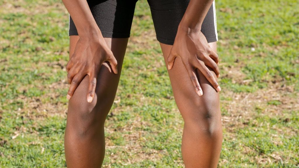
How Misaligned Feet Affect Knees
Misaligned feet can disrupt normal foot position, altering the way forces move up the leg and into the knee. As the base of the body, feet play a crucial role; when one is out of alignment, load paths through the ankle, tibia, and femur adjust. This shifted burden increases strain on certain knee compartments, amplifies friction across cartilage, and may accelerate degeneration that leads to osteoarthritis. Notably, foot deformities can cause ongoing knee pain even when X-rays or scans appear normal, as the changed mechanics induce pain and inflammation before structural changes are visible on imaging.
1. Overpronation Impact
Overpronation is when your foot rolls inward too much while walking or running, leading to bad foot alignment. If your foot overpronates, the tibia internally rotates, and the knee follows more towards the middle, loading the medial compartment and predisposing you to medial tibiofemoral osteoarthritis over the years of repeated stress. This altered foot posture increases pressure on tibiofemoral cartilage, accelerating wear and potentially causing joint discomfort. Overpronation commonly causes pes anserine tendinitis and frequent knee pain as the pes anserinus insertion is overloaded by the altered pull of adjacent muscles and tendons.
2. Supination Strain
Supination, characterized by poor foot alignment, occurs when the foot rolls too far outward, placing weight on the outside edge. This abnormal foot posture shifts stress to the outside of the knee, potentially leading to frequent knee pain along the lateral joint line. Additionally, supination minimizes shock absorption, compelling impact to travel up a less compliant chain, which induces an uneven gait and balance problems that amplify instability. Over time, the lateral structures bear excess load, increasing the risk for knee osteoarthritis due to joint degeneration.
3. Flat Feet Pressure
Flat feet result in a loss of natural arch support and a flatter-than-normal plantar surface during stance. This correct foot posture can lead to a loss of foot stability, causing more load to shift to the knee, ultimately affecting the knee function and pressure distribution across the joint. Flat arches are associated with an increased incidence of medial meniscus damage and knee osteoarthritis, particularly because the medial side experiences recurrent high contact stress. Supportive shoes or custom orthotics that provide arch support can help alleviate knee pain by altering the direction of forces and improving overall foot alignment.
4. High Arch Tension
High arches generate too much pull on the outside of the foot and the outer knee and ankle. Decreased shock absorption with cavus feet makes each step transmit greater impact forces to the knee, which increases cartilage strain and can lead to joint discomfort. High arches are frequently accompanied by tight calves and a susceptibility to Achilles tendonitis, which changes ankle mechanics and the kinetic chain. Checking arch height during any knee pain workup is important, as correcting foot posture can reduce joint load and restore an effective walking method.
Beyond The Obvious Signs
Foot misalignment can start small and lurk behind otherwise normal appearing knees, leading to issues like knee varus and frequent knee pain. You must look beyond obvious signs, as early knee osteoarthritis often manifests as subtle changes in foot posture and gait before pain, swelling, or imaging abnormalities are present. Recognizing these warning signs early provides you with the opportunity to alter loading patterns and decelerate joint degradation.
Uneven Wear
Uneven wear on your shoes can often be indicative of how forces are traveling from your foot to your knee. When the lateral edge of the sole wears faster, you could be supinating and loading medially through the knee. When the inner edge wears faster, pronation can increase valgus forces at the knee. A worn heel bulge on one side might mirror a leg-length difference that torques pelvis and knee mechanics. These patterns are not a diagnosis, but they are cheap, available clues you can monitor over months.
Wear type | Typical foot action | Likely knee/hip implication |
Outer sole wear (lateral) | Supination, under-pronation | Increased medial knee load, possible inner knee OA |
Inner sole wear (medial) | Over-pronation | Increased knee valgus stress, lateral hip strain |
Heel-center wear | Neutral gait | Balanced loading, less directional risk |
Heel offset on one shoe | Asymmetry, leg-length difference | Pelvic tilt, uneven knee joint stress |
Take pictures of your shoes and mark them every 2 to 3 months to see if they have any excessive wear. Left versus right. That straightforward table assists you and any clinician to develop a plan that focuses on the specific biomechanical problem.
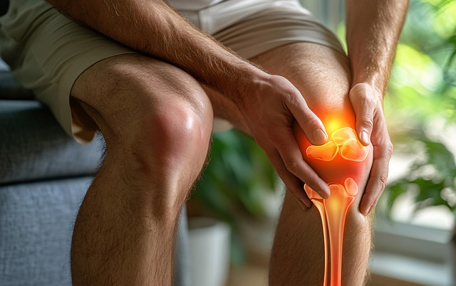
Silent Progression
Knee osteoarthritis can progress silently for years before you experience acute pain. Cartilage thins and bone adapts without overt inflammatory signs. Many people live with knee pain for years while X-rays read surprisingly normal because early cartilage loss often doesn’t register on standard images.
Small shifts in foot alignment, uneven pronation, supination, or slight leg-length discrepancy can alter joint loading sufficiently to initiate this cascade. Science says adjusting foot angle during walking reduces pain. One study observed a 2.5-point reduction on a 10-point pain scale, comparable to typical OTC pain medications. This means easy gait tweaks and tailored strategies can make a difference. Monitor range of motion, joint stiffness after rest, and ability to climb stairs. These functional checks catch decline earlier than waiting for swelling.
Postural Clues
Observe your own posture and gait. Subtle postural signs come before obvious knee trouble and indicate where to intervene.
- Knee valgus (knees caving in) during a squat or jump landing.
- Varus knee (bowlegs) with increased load on the inner knee.
- Hip drop on the swing side indicates weak hip abductors that change knee mechanics.
- A pelvic tilt or one shoulder lower than the other indicates leg-length discrepancy.
- Asymmetric toe-out or toe-in with gait demonstrates compensatory foot angle shifts.
Be sure to observe both barefoot and shod and record a quick video for side-by-side comparison over the course of months. Tailored tests trump generic solutions.
Diagnosing The Root Cause
Only a careful, systematic procedure can discover the cause of your knee pain. Start by seeing the big picture: foot alignment, knee biomechanics, muscle function, and imaging. Converge objective gait data, hands-on tests, and targeted scans. Log it all in a straightforward template so discoveries regarding foot posture direct treatment, and allow you to monitor progress.
Gait Analysis
Conduct an analysis using a gait lab or treadmill with motion capture to research your walking and running patterns. Capture stride length, cadence, foot progression angle, and limb-force lines under load. Video and force-plate data expose regularities that plain observation overlooks. Custom gait retraining can then focus specifically on the pattern that overloads your knee.
- Excessive knee adduction moment means the knee bucks inward, over and over, increasing medial joint load, and is associated with medial knee osteoarthritis. Retraining and orthoses can decrease this load.
- Out-toeing (external foot progression) — rotates knee mechanics and changes load patterns. It can be due to hip rotation or foot alignment problems and can be corrected with gait cues.
- In-toeing (internal foot progression) directs stress onto different cartilage regions and is frequently associated with femoral or tibial torsion. Physical therapy and gait retraining aid.
- Stride length is shortened over heightened cadence, which pumps up impact rate and potentially speeds wear. Interventions with strength work and step-length cues.
- Asymmetrical loading — one limb absorbs more load, typically from limb-avoidance related pain or leg length difference. Fixing symmetry minimizes one-sided wear and tear.
- Late heel-off or early forefoot loading alters the timing of force through the knee and ankle and is occasionally associated with tight calves or forefoot pathology.
Document these anomalies and indicate which ones shift with straightforward signals in your chart.
Physical Assessment
Diagnosing the root cause of knee issues involves evaluating hip and ankle range, as they significantly impact knee movement and overall foot alignment. It’s essential to identify weak hip abductors or a tight IT band that can alter knee tracking and contribute to conditions like medial compartment knee osteoarthritis. Additionally, local foot problems such as flatfoot or high arches, bunions, and heel pain can refer pain to the knees or change load paths, ultimately affecting joint function.
Testing muscle strength and passive range of motion is crucial. Specific provocation tests should be used to reproduce knee symptoms while altering foot posture. Documenting findings in a structured sheet that includes static alignment, dynamic deficits, and provoked symptoms will inform whether foot correction, strengthening, or gait retraining will likely provide relief and improve walking patterns.
This structured log serves as a baseline for follow-up and helps determine the effectiveness of interventions. Addressing foot deformities and ensuring correct foot posture can significantly enhance knee function, especially in osteoarthritis patients experiencing ongoing knee pain. By focusing on limb alignment and the natural alignment of the feet, we can develop effective walking methods that alleviate discomfort.
Imaging Techniques
MRI and specific radiographs provide additional information. MRI reveals cartilage health, bone marrow lesions, and soft-tissue status that X-rays don’t see, which is important because lots of people have persistent knee pain, even with normal X-rays. They are great for diagnosing the root cause.
Modality | What it shows | When to use |
Weight-bearing X-ray | Joint-space narrowing, osteophytes, alignment | Initial structural staging |
MRI | Cartilage, meniscus, bone marrow lesions, soft tissue | Early degeneration, treatment tracking |
Lateral radiograph | Anteroposterior alignment detail | Compare with frontal views to assess malalignment |
Compare images over time to monitor the impact of the intervention. Don’t use old-school biomechanical models. Certain podiatric approaches from the 1970s overlook dynamic patterns you see in gait labs. Combine the imaging with the gait and physical data for a crisp, actionable diagnosis.
Proactive Management Strategies
Misaligned feet can lead to bad foot alignment, altering the path of load traveling up the leg and potentially increasing stress to the knee joint. You need a plan that realigns, minimizes peak loads, and creates muscular support. The steps below detail actionable, research-backed strategies to maintain proper foot posture and decelerate joint destruction while reducing symptoms.
Supportive Footwear
Opt for rearfoot-stabilizing and arch-supportive shoes to control excessive pronation or supination, which changes the way the knee loads. Firm heel counters lock the heel in place and limit torsion up through the tibia, and proper arch support helps spread pressure across the foot instead of directing it into either the medial or lateral compartments of the knee. Bad shoes, flat, worn-out soles, overly flexible heels, or narrow toe boxes will exacerbate misalignment and magnify knee pain.
Pick footwear with these features:
- Firm heel counter for rearfoot control
- Structured midsole for stability
- Adequate arch support matched to your foot type
- Shock-absorbing cushioning in the heel and forefoot
- Wide, stable base and appropriate toe-box width
- Durable outsole with grip to prevent slips
- Replace shoes every 6–12 months or when the midsole compresses
Custom Orthotics
If you have stubborn foot malalignment, custom insoles correct your individual mechanics better than generic inserts, so think about adding them. A customized orthotic transfers plantar loading, unloads affected knee compartments, and can modify tibial rotation forces that impact cruciate and patellofemoral contact. Devices range from full custom molded shells to targeted support such as shelf insoles or the ergo pad redux heel for localized cushioning and rearfoot control.
Use orthotics to:
- Rebalance pressure under the foot
- Control excessive pronation or supination
- Improve knee alignment during gait
- Reduce painful hotspots and swelling
Follow up by tracking pain scores, distance walked, and joint range. Modify or switch devices if pain doesn’t fall or if function doesn’t improve.
Targeted Exercises
Strength and flexibility modifications change biomechanics. Build intrinsic foot muscles, calves, quads, and hamstrings to absorb shock and stabilize joints. Stretch tight calves and hamstrings. These tight muscles limit ankle dorsiflexion and promote compensatory knee motions. Include some mobility drills to keep the ankle-knee-hip chain moving in coordinated patterns.
- Toe curls: Sit and curl toes around a towel for 3 sets of 10. This exercise develops intrinsic foot strength and enhances arch control.
- Golf ball roll: Roll the ball under the arch for 2 to 3 minutes. This minimizes plantar tight spots and enhances proprioceptive feedback.
- Ankle mobility drill: Kneel with foot flat, shift knee forward to the wall without heel lift. Perform three sets of ten repetitions per side. This regains the dorsiflexion required for normal walking.
- Calf raises and eccentric hamstring slides: 3 sets of 12 each. Fortify push-off and govern limb deceleration.
Personalize these plans: Small changes to foot angle while walking can cut maximum knee loading by about 4% versus an increase of 3% if you make no change. Customize orthotics, footwear, and gait cues to your specific pattern, which frequently equals or outperforms the impact of basic pain meds and can put off surgery.
Lifestyle And Long-Term Health
Misaligned feet alter the forces that travel through your knee over time, so lifestyle choices directly determine how fast cartilage wears away and how much pain you experience. Start with weight: carrying extra mass raises joint load with each step, and losing weight reduces that load in a clear, measurable way. For every 0.45 kg (1 lb) you drop, you take approximately 4 kg (8 lbs) of pressure off of weight-bearing joints. For someone with early medial knee osteoarthritis, a 5 to 10 percent bodyweight loss can reduce joint stress enough to reduce cartilage loss and relieve symptoms.
Be active, and choose your activities carefully. Low-impact exercise, such as biking, swimming, elliptical work, and walking briskly, keeps cartilage fed and muscles powerful without the pounding that aggravates alignment issues. Strength training of the quadriceps, hip abductors, and calf musculature controls knee tracking and combats the abnormal forces caused by foot malalignment. If you already have knee pain, aim for consistency, not intensity. Research demonstrates that people with knee osteoarthritis walk around 10.8 fewer steps per minute than healthy counterparts, so increasing daily step count is more important than fast bursts of effort.
Don’t take long periods sitting or standing on uneven floors. Sitting too long decreases joint lubrication and leads to stiffness that transfers load onto smaller areas of cartilage when you move again. Standing on inclined or uneven surfaces induces compensatory foot and knee angles that can focus wear and tear on the inner knee compartment. When work or travel necessitates extended standing, utilize mini shifts, mini walks, or mats to distribute the load and minimize repetitive stress.
Lifestyle and long-term health. Shoes designed to address over-pronation or supination can alter knee moments and reduce peak loading. Personalized adjustments matter: research suggests tailoring foot angle and alignment to your unique gait often works better than one-size-fits-all devices. Gait retraining — altering your walking pattern — can decrease maximum knee load by approximately 4% and drop pain scores by approximately 2.5 points on a 10-point scale. It is intricate. It generally takes approximately six weeks with regular practice to learn new gait patterns, and long-term follow-up is necessary to observe if individuals maintain those modifications. Experts caution that real-world viability differs, so combine retraining with strength work and shoe changes.
Expect a multi-part plan: lose weight where needed, build strength, choose low-impact exercise, avoid harmful postures, and use footwear or orthotics that fit your biomechanics. Track steps, pain, and function. If gait work is recommended, anticipate a minimum of six weeks of directed, repeated practice and occasional readjustment.
Conclusion
You can now see how foot alignment can change the load on the knee. Minor changes in foot angle increase stress to cartilage by obvious, quantifiable levels. Gait analysis and simple strength tests indicate where the issue originates. These targeted changes, including orthotics, shoe choice, and hip and ankle work, combined with a clear rehab plan, can cut pain and slow cartilage loss. Test a course of an OTC arch support for four to six weeks. Monitor pain, steps taken, and stair climbing experience. If pain persists or escalates, obtain a professional consultation and imaging. Real gains come from steady work and small wins: a few daily hip moves, smarter shoes, and better step habits. Begin with a single change this week and feel the difference.
Frequently Asked Questions
1. Can Misaligned Feet Really Speed Up Knee Arthritis?
Yes. Misalignment of the feet, or bad foot alignment, alters your gait, adding additional strain to knee cartilage and joint structures, contributing to arthritis progression over time.
2. What Foot Problems Most Affect Knee Health?
Flat feet (overpronation) and high arches (oversupination) affect foot alignment, changing knee loading and increasing the risk of joint wear.
3. How Will I Know If My Knee Pain Comes From My Feet?
Look for uneven shoe wear, ankle or calf pain, and knee pain that varies with footwear, which may indicate issues like bad foot alignment or foot deformities. A gait or biomechanical assessment confirms the link.
4. Will Orthotics Or Shoe Changes Stop Arthritis Progression?
Custom orthotics and supportive shoes can enhance foot alignment, minimizing abnormal knee load and providing relief for osteoarthritis patients, easing pain and slowing progression.
5. What Exercises Help If My Feet Are Misaligned?
Strengthening hip stabilizers, calf muscles, and foot intrinsic muscles helps improve foot alignment. Balance and mobility exercises optimize your gait and minimize joint stress.
6. When Should I See A Specialist About Foot-Related Knee Pain?
See a physical therapist or orthopedist if pain restricts activities, lingers for weeks, or you observe visible gait changes related to foot alignment. Early evaluation aids results.
7. Can Lifestyle Changes Reduce My Arthritis Risk Even With Misaligned Feet?
Yes. These strategies can reduce knee stress and slow the progression of arthritis, including maintaining proper foot alignment and using appropriate footwear for effective walking methods.
Find Real Relief From Chronic Knee Pain By Fixing Your Foot Alignment
Chronic knee pain has a way of creeping into everything. Walking, working, even just getting through the day can start to feel like a chore. What most people don’t realize is this: your knees might not be the real problem. When your feet are out of alignment, every step sends extra stress up into your knees, and over time, that adds up to pain that won’t go away.
At The Shoe Doctor, we take a different approach. Instead of chasing symptoms, we look at how your body moves from the ground up. Using advanced 3D foot-mapping technology, we pinpoint exactly where your alignment is breaking down, how your gait is affecting your knees, and where pressure is building with every step.
From there, we create custom orthotics designed specifically for your feet and your movement. The goal is simple: restore proper alignment, reduce strain on your knees, and help your body move the way it was meant to. When your foundation is right, everything above it starts to improve.
With over 20 years of experience, Russell has helped countless people get out of chronic pain and back to living normally again. Working alongside Spine and Injury Medical Center, we also look at posture and full-body mechanics so your results last, not just for now, but long term.
If knee pain has been limiting you, don’t settle for temporary fixes. Get to the source of the problem and finally feel the difference. Book your free consultation today and take the first step toward moving better, feeling stronger, and living without constant knee pain.
Disclaimer
The materials available on this website are for informational and entertainment purposes only and are not intended to provide medical advice. You should contact your doctor for advice concerning any particular issue or problem. You should not act or refrain from acting based on any content included in this site without seeking medical or other professional advice. The information presented on this website may not reflect the most current medical developments. No action should be taken in reliance on the information contained on this website, and we disclaim all liability for actions taken or not taken based on any or all of the contents of this site to the fullest extent permitted by law.