Key Takeaways
- Flat feet alter your foot biomechanics and heighten knee strain by facilitating overpronation and internal tibial rotation, increasing the likelihood of medial knee cartilage injury and osteoarthritis. Have a clinician check alignment early before it progresses.
- Collapsed arches and ankle overpronation shift your weight inward and transform the tibiofemoral angle, resulting in knee valgus and patellar tracking issues that often manifest as medial or anterior knee pain. Measure arch height and gait to inform treatment.
- A thorough diagnosis should consist of a directed physical exam, gait analysis with pressure mapping if possible, and focused imaging like X-ray or MRI to detect cartilage damage and malalignment. Document findings with standardized tools to track change.
- Early multimodal management reduces long-term damage. Use supportive footwear or custom orthotics to restore alignment. Follow a tailored exercise program to strengthen foot, hip, and thigh muscles. Incorporate balance training to correct compensatory movement.
- Addressable risk factors such as excess body mass and footwear have huge effects on knee load and symptom severity. Emphasize weight management and supportive shoes as two concrete things you can do today.
- Keep in mind that age, genetics, occupational demands, and systemic conditions contribute to symptoms, so approach holistically and co-manage with specialists when pain lingers or function worsens.
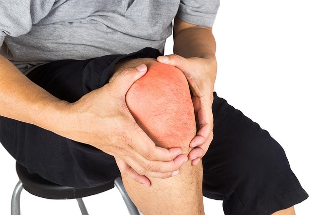
Do flat feet cause chronic knee pain? Flat feet frequently alter your foot’s strike and the transmission of force up your leg, which can increase pressure on knee joints and tendons. You might feel a dull ache at the front or side of the knee after walking, standing, or light running. Specifically, excess tibial rotation and reduced shock absorption increase load on the patellofemoral joint and medial structures by quantifiable amounts. Basic screen tests, such as observing gait and taking an arch height index, can help correlate foot posture to symptoms. Conservative measures like supportive insoles, targeted strengthening of tibialis posterior and glute medius, and gait retraining reduce knee load and reduce pain in most people.
The Biomechanical Link
Flat feet alter the weight bearing of your foot and the transmission of forces up your leg, impacting knee function and increasing the risk of specific knee cartilage damage. This text deciphers the biomechanical domino effect from foot to knee, explaining what changes, why it stings, and how this increases danger for chronic knee pain.
1. Collapsed Arches
These are cases of fallen arches – falling arch height and falling support from the foot. With less arch, your foot flattens under load, causing your body weight to shift more medially, which increases pressure on the inner (medial) knee compartment and can accelerate specific knee cartilage damage. Over time, this inward shift raises the risk of medial tibiofemoral cartilage injury approximately 1.3 to 1.4 times relative to normal arches. Symptoms you’ll observe are foot soreness after standing, frequent knee pain when walking or on stairs, and an altered gait pattern. Gait analysis frequently reveals these dynamic changes that a static exam overlooks. Morning stiffness in the knees that subsides with movement can indicate overnight rest, eliminating inflammation created by daytime biomechanical factors.
2. Ankle Pronation
Ankle pronation, the hyperdorsal flexion of the foot during stance, is often associated with flat feet and can lead to specific knee cartilage damage. When your ankle overpronates, your tibia and femur do as well, creating strange knee movement that increases load on the medial compartment of the knee OA. This motion strains ligaments and tendons surrounding the joint, decreasing knee function and stability chronically, aggravating frequent knee pain, and increasing the risk of degenerative change. Prolonged pronation makes your hips and lower back compensate, potentially leading to hip pain from overcompensation in your upper leg or low back pain from altered posture and body imbalance.
3. Tibial Rotation
Tibial rotation is the twisting of the shin bone caused by abnormal foot mechanics in flat feet, which can lead to frequent knee pain. Internal tibial rotation stresses the medial knee compartment more severely and alters contact patterns on cartilage, increasing the risk of osteoarthritis outcomes. This rotational stress is directly connected to knee malalignment and may contribute to specific knee cartilage damage, resulting in meniscal tears or ligament strain when the knee is exposed to repeated non-neutral positions. Fixing tibial rotation involves addressing foot posture, hip control, and movement patterns simultaneously.
4. Knee Valgus
Knee valgus, characterized by the inward angling of the knees, often correlates with flat foot posture and can lead to medial compartment knee OA. This condition applies disproportionate force to different knee compartments, accelerating specific knee cartilage damage and increasing your susceptibility to frequent knee pain, particularly in those with poor arch support.
5. Patellar Misalignment
Flat feet can alter how the patella tracks in the femoral groove, generating lateral patellar pain and increased susceptibility to specific knee cartilage damage. This misalignment causes joint stiffness and leads to decreased knee function and anterior knee pain during activities such as squats, stairs, and standing for long periods. Strengthening hip muscles and enhancing neuromuscular control effectively reorient these forces to unburden the knee.
Identifying The Symptoms
Flat feet alter the biomechanics of your lower limb, and that alteration manifests itself as a predictable cluster of symptoms you can monitor. Notice the patterns, not the one-off twinges. Typical symptoms are persistent pain in your knees, feet, and lower legs, which is usually worse after running, walking long distances, or climbing stairs. Activity-linked pain is typical. You may be fine at rest, then notice aching or sharp pain during and after exercise. Among a group of 1,903 flat-footed patients, 22% experienced knee pain on most days, highlighting just how frequently symptoms become chronic if left untreated.
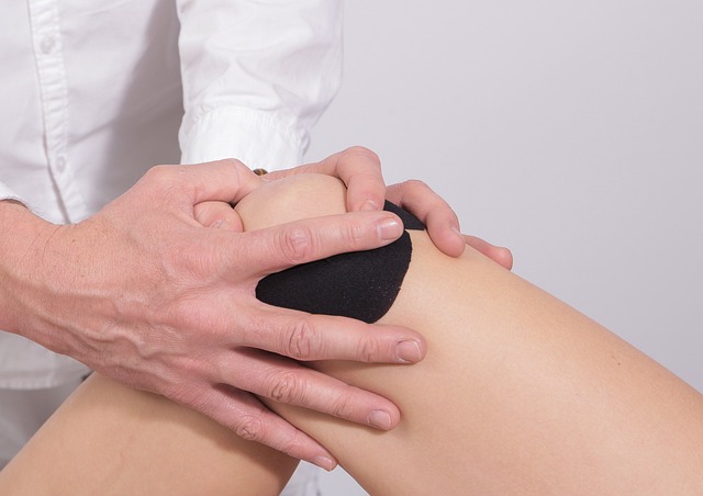
Joint tenderness, swelling, and limited range of motion of the knee are significant warning signs. Tender areas are sore to the touch along the joint line, and swelling can give your knee a puffy appearance or stiffness. Limited range of motion presents as difficulty fully extending or flexing your knee, or a feeling that the joint is ‘stiff’ after sitting. Morning stiffness in your back or knees that abates with activity may indicate nighttime repair from biomechanically induced inflammation during the previous day. If stiffness gets better once you’ve been moving around for ten to twenty minutes, that pattern indicates load-related inflammation, not a singular injury.
Anticipate concomitant symptoms in surrounding areas since the foot establishes the chain for the remainder of the limb. Flat feet tend to generate foot complaints like arch fatigue, heel pain, or generalized soreness through the sole. Hip aches and low back pain occur when your body compensates. Overpronation, where the ankle rolls inward, can cause the knee and hip to rotate inward, changing muscle and joint loads. This inward rotation raises your risk. People with flat feet have 1.3 times the odds of knee pain and 1.4 times the odds of knee cartilage damage, which speaks to a systemic effect not isolated to the foot.
Weakness in the foot muscles manifests as a loss of arch height and is directly connected to achy knees. If your intrinsic foot muscles fail to prop up the arch, the remaining leg is overloaded. Identify the symptoms.
Record symptom onset, duration, and severity to note trends and responses to treatment. Observe when pain begins in relation to activity, how long it persists, if it wakes you at night, and if easy measures like shoes or rest alleviate it. Use a simple log: date, activity, pain score from 0 to 10, location, and what eased or worsened it. Over time, you will observe if the interventions assist.
Running and stair climbing are two activities that often trigger symptoms in flat-footed individuals because both add load and require foot stability. Remember that our feet dictate whole-body movement, so it is a major key in preventing and treating chronic knee pain.
The Kinetic Chain Reaction
Flat feet significantly alter how your body first hits the ground, and this alteration courses upward through joints and soft tissues, affecting knee function. When your foot pronates excessively, the tibia and femur can rotate inward, leading to altered knee measures and increasing the risk of knee pain. This part deconstructs those connections so you can understand how an issue at the foot can cause long-term knee pain and associated back symptoms.
Hip Imbalances
Flat feet can significantly alter your gait, leading to issues such as frequent knee pain. When your foot pronates, the hip must compensate to maintain balance, which may result in weak gluteus medius and maximus muscles due to inefficient loading. This imbalance can also cause the tensor fasciae latae to become tight and overactive. Such weak or tight hip muscles allow for inward rotation of the femur, which disrupts knee function and increases shear and compressive stresses on the patellofemoral joint and tibiofemoral cartilage. Research has shown a connection between flat feet and a higher risk of specific knee cartilage damage, emphasizing the importance of addressing these biomechanical factors.
Moreover, hip imbalances can affect loading on the lumbar spine, leading to lower back strain when the hips fail to stabilize the pelvis. By focusing on strengthening hip muscles and enhancing flexibility through targeted exercises, we can improve knee health and mitigate the long-term risks of knee tissue damage.
Pelvic Tilt
Pelvic tilt refers to the forward or backward tilt of the pelvis in relation to your spine. Changed foot and leg mechanics push the pelvis into a new resting position. An anterior tilt enhances lumbar lordosis and alters the mechanical axis through the hip and knee, while a posterior tilt flattens the lumbar curve and shifts load patterns. Either tilt can amplify forces at the knee by displacing the line of force from the joint’s center.
When you have flat feet, repeated inward rotation of the lower limb over time can promote a compensatory pelvic tilt, which then feeds back to worsen knee function. Assessing pelvic alignment is essential in a thorough knee pain plan. Manual assessment, functional tests, and gait analysis reveal these shifts. Treating only the knee without addressing the pelvis leaves the underlying mechanical axis uncorrected.
Lower Back Strain
Lower back strain is a frequent guest in those with stubborn foot and knee problems. As foot pronation alters gait, lumbar spine mechanics change, perhaps causing the spine to flex or rotate more with each step, increasing cumulative load on intervertebral discs and facet joints. You may experience morning stiffness in your back or knees that dissipates with movement. This can mirror the overnight decrease of inflammation due to daytime biomechanical strain.
About the Kinetic Chain Reaction, gait analysis reveals those dynamic compensations that static checks overlook, demonstrating how aberrant foot motion transmits forces to the lumbar region. Correcting foot posture with orthotics, specific exercises, and gait retraining decreases aberrant spinal loading and minimizes the risk of chronic lower back strain.
|
Foot problem |
Knee symptoms |
Back/hip symptoms |
|
Overpronation / fallen arch |
Medial knee pain, patellofemoral pain, cartilage wear |
Increased lumbar strain, hip internal rotation |
|
Excessive tibial rotation |
Anterior knee pain, instability |
Pelvic tilt, low back stiffness |
|
Long-standing flat feet |
Chronic knee osteoarthritis risk |
Persistent lower back pain, altered gait |
Seeking A Diagnosis
Getting a diagnosis for persistent knee pain and flat feet isn’t always immediate. It takes multiple visits and time, especially when assessing specific knee cartilage damage and foot conditions. It introduces into the procedure actionable steps that help to demystify the cause and direct treatment. It can provoke stress, waiting, and disparities in access to specialists and diagnostic tools. Here’s a targeted roadmap you can apply to go from symptom to particulars, with instruments and metrics that enhance precision and monitor change over time.
- Physical exam, gait analysis, and imaging tests.
- Identify knee cartilage damage and foot morphology.
- Take advantage of knee pain rating scales and foot-arch measuring devices to track your progress.
Physical Examination
A clinical exam begins with assessing foot arch height, alignment, and flexibility. You need the arch measured in both weight-bearing and non-weight-bearing positions to see functional collapse. Observe static foot posture and look for pes planus, forefoot abduction, and heel valgus. Perform the navicular drop test and a simple footprint or arch index. Knee posture assessment follows. Inspect valgus or varus alignment, measure Q-angle, and note asymmetry between limbs.
Palpate for joint line tenderness, patellar tracking, and localized swelling. Test active and passive range of motion at the knee and ankle, documenting degrees of flexion and extension, crepitus, or locking. Check ligament stability with drawer tests, varus/valgus stress tests, and test tendon integrity, especially patellar and Achilles tendons, with resisted contraction and palpation. Record everything in a checklist format so you can contrast subsequent visits and talk clearly to other clinicians.
Gait Analysis
Gait analysis examines walking and running mechanics to identify abnormal foot and knee movement that may be fueling frequent knee pain. It scrutinizes stance and swing phases, the timing of pronation and heel rise, and looks for early or prolonged pronation that exerts torque on the tibia and knee. Digital pedobarography measures dynamic pressure distribution under the foot, quantifying pronation, contact area, and peak pressure zones, which are crucial for assessing foot morphology. These data reveal where the load concentrates and when, helping to understand knee function better.
Gait evaluation also uncovers compensatory strategies such as hip drop, external rotation of the leg, or excessive lumbar motion that may place stress on the knee and back. Employ video capture at various speeds and, if available, 3D motion analysis to correlate kinematics with symptoms. A table below outlines typical gait abnormalities and anticipated knee consequences of each, including specific knee cartilage damage assessments.
|
Gait abnormality |
Typical foot sign |
Impact on the knee |
|
Prolonged pronation |
Medial pressure, collapsed arch |
Increased tibial internal rotation, patellofemoral pain |
|
Early heel rise |
Forefoot loading |
Anterior knee stress, reduced shock absorption |
|
Excessive supination |
Lateral pressure |
Lateral compartment overload, iliotibial band tension |
Imaging Tests
Standard imaging incorporates weight-bearing X-rays, MRI, and long limb alignment radiographs to evaluate bone alignment and joint space. MRI demonstrates tibiofemoral cartilage damage, patellofemoral changes, meniscal lesions, and early osteoarthritis overlooked by plain films. Long limb films allow us to quantify varus or valgus malalignment that connects foot mechanics to knee load.
Foot imaging—weight-bearing foot X-rays or CT—illuminates arch collapse, talar displacement, and deformity severity. Periodic imaging is needed to monitor the progression of knee osteoarthritis and foot deformities if symptoms worsen or conservative care fails. Testing may be limited by access and expense, so select tests according to the degree of symptomatology, loss of function, and clinical findings.
Proactive Management Plan
A proactive management plan minimizes the likelihood that flat feet will induce progressive knee cartilage damage, assessments, and chronic pain. Early intervention focuses on realignment, load distribution, and muscular support to prevent altered gait patterns that increase medial compartment knee OA or lateral tibiofemoral stress. Here are actionable, research-based steps you can implement, along with a convenient checklist to keep you on track.
Supportive Footwear
Select shoes that provide arch support, cushioning, and lateral stability to control overpronation, as these features are essential for maintaining optimal knee function. Athletic shoes designed for overpronation often include internal medial posts or stiffer midsole material to restrict excessive inward roll and assist in reestablishing normal tibial rotation during stance. Good shoes minimize the sharp spikes of force that go through your knee, helping to reduce frequent knee pain and potentially slow down knee cartilage damage assessments.
When searching for the right footwear, find a shoe with a firm heel counter to hold your rearfoot in place, a wide toe box to allow forefoot splay, and a sole with controlled flexion that bends at the forefoot instead of the midfoot. Features like a rocker sole or foam layers for shock absorption are handy if you experience walking pain. Test multiple pairs while standing and walking to ensure they seem stable and prevent excessive pronation, which can lead to medial compartment knee OA.
Custom Orthotics
Custom orthotics offer personalized arch support and realignment according to your specific foot structure and walking pattern. They shift plantar pressure, taking load off overloaded areas and unloading knee compartments and cartilage. This is particularly important if gait analysis reveals an excessive amount of pronation.
Orthoses are valuable for managing severe pes planus and for patients at risk of knee osteoarthritis progression. Expect an initial fitting and follow-up adjustments. Foot morphology and activity levels change, so periodic reassessment ensures the orthotic continues to help. Combine orthotics with supportive shoes for the best results.
Targeted Exercises
Fortify the foot intrinsics, tibialis posterior, calf complex, quads, and hip abductors to assist with alignment from foot to pelvis. Basic drills, such as lifting the four little toes while keeping the big toe down, help to strengthen intrinsics and arch control. Calf raises, resisted ankle inversion, single-leg squats, clamshells, and hip bridges increase the kinetic chain that shields the knee.
Add in daily stretching of your gastrocnemius, soleus, hamstring, and IT band to alleviate compensatory tightness. Supplement with balance and proprioception drills, such as single-leg stance and wobble-board work, to enhance dynamic stability and gait. Structure a weekly plan with three strength sessions, two mobility sessions, and daily short balance drills tailored to your severity and goals.
Weight Management
Excess BMI adds compressive load to the knee and hastens cartilage attrition. Losing even 5 to 10 percent of body weight can reduce pain and slow osteoarthritis. If you are overweight, make sure you follow a calorie-controlled diet and low-impact aerobic activity such as cycling, swimming, or walking to safeguard your feet while shedding pounds.
Monitor weight and knee pain scores weekly to observe the impact of your modifications. Intermingle weight targets with shoes, orthotics, and exercise compliance for a quantifiable, multi-pronged plan.
Checklist for adherence: Get a gait analysis, choose supportive shoes, use custom orthotics as prescribed, follow a weekly exercise plan, monitor weight and pain, and schedule reassessments every 3 to 6 months.
The Unseen Influences
Flat feet alter the way forces are transmitted through your lower extremity. With your foot collapsing in, your ankle overpronates, and your tibia internally rotates. This rotation disrupts knee alignment and load distribution, potentially leading to frequent knee pain or heaviness in your knees or ankles after extended walking, standing, running, or stair climbing. The body operates as one system: poor foot position often manifests as altered knee function, uneven cartilage stress, and localized inflammation that may worsen after activity yet feel better after a night’s rest.
Your genetics, age, and lifestyle determine how intense your symptoms get. If you have more pliable arches, you might be susceptible to higher collapse. As we age, our tendons and ligaments become less elastic, and therefore the same mechanics create more strain. If you stand for long periods or do repetitive impact activities, your tissues receive repetitive microstress. A study of 1,903 flat-footed people revealed that just 22 percent had daily knee pain. Over half had medial or lateral knee cartilage damage in the absence of consistent pain, which demonstrates that structural change can outpace what you feel.
Job requirements and shoes impact symptom formation in obvious ways. Standing or floor-to-knee jobs add cumulative load to your knee. Shoes with no arch support allow the foot to overpronate even more. Thin-soled or worn-out shoes do not absorb as much shock, which increases force transmission up the leg. If you run or climb stairs, you are likely to experience more pain because those activities increase peak joint forces and demand more pronation control upon push-off and impact.
Underlying musculoskeletal or systemic diseases complicate management. Conditions like inflammatory arthritis, including rheumatoid arthritis, can introduce synovial inflammation that amplifies pain from biomechanical stress. Tendinopathies, past ligament injuries, and malunited fractures alter local mechanics, potentially diminishing the knee’s ability to withstand the altered loading caused by flat feet. Symptoms such as morning stiffness that improve with activity may indicate inflammation subsiding overnight, while biomechanical irritants can escalate pain and swelling during the day.
Consider these less obvious factors for clinical or personal evaluation. Important elements to assess include shoe wear and age, work and training load, body mass index, hip and ankle range of motion, previous lower limb injuries or surgeries, systemic inflammatory symptoms, and pain patterns associated with specific tasks like running or stair climbing. Monitor the timing of symptoms, whether stiffness improves with activity, and any functional limitations. Utilize this data to inform interventions: footwear modifications, activity adjustments, orthotics, targeted strengthening, or referrals for imaging if structural damage is suspected.
Conclusion
Flat feet can cause chronic knee pain via obvious biomechanical alterations in foot dynamics and leg weight bearing. You experience strain at the knee when the arch collapses, the foot rolls inward, and the thigh rotates. Simple tests and a quick exam reveal the problem. Targeted fixes provide real relief. Go for arch-support insoles, pick shoes with firm midsoles, target hip and ankle strength, and include motion-control drills. Apply a slow load increase to your training. Monitor pain and adapt accordingly. If pain persists or worsens, obtain a specialist consultation and imaging if necessary. Knee pain relief, ready? Begin with a biweekly shoes, pain, and step log.
Frequently Asked Questions
1. Can Flat Feet Directly Cause Chronic Knee Pain?
Yes. Flat feet can alter the way your knee functions and absorbs shock. Over time, this can lead to specific knee cartilage damage and chronic knee pain if left untreated.
2. How Will I Know If My Knee Pain Is Linked To Flat Feet?
Watch for ankles that roll inward, frequent knee pain after walking or running, and discomfort on the front or inside of the knee. A clinician can verify knee function with a gait exam and images if necessary.
3. Will Orthotics Stop My Knee Pain From Flat Feet?
Custom or OTC orthotics often diminish abnormal foot motion and alleviate knee stress, particularly in cases of knee malalignment and frequent knee pain. They work for a lot of people, but not all. Wear them to work out with a professional opinion!
4. Should I See A Specialist For Flat-Foot-Related Knee Pain?
Yes. Visit a PT, podiatrist, or orthopedist to identify the source of frequent knee pain and prescribe specific exercises and treatments to avert permanent damage.
5. Can Exercises Fix Flat Feet And Reduce Knee Pain?
These targeted strengthening and mobility exercises enhanced both foot arch support and knee function, effectively alleviating frequent knee pain and decreasing re-injury risk if performed regularly.
6. When Is Surgery Necessary For Flat Feet And Knee Pain?
Surgery is uncommon and typically reserved for cases of severe structural deformity or joint damage, particularly when knee function is significantly impaired after months of conservative care.
7. What Else Can I Do Right Now To Reduce Knee Pain From Flat Feet?
Begin with supportive shoes, temporary foot orthotics, pain-soothing ice, and low-impact exercise to improve knee function. Schedule a professional analysis to craft an enduring strategy.
Find Real Relief From Chronic Knee Pain By Fixing Your Foot Alignment
Chronic knee pain has a way of creeping into everything. Walking, working, even just getting through the day can start to feel like a chore. What most people don’t realize is this: your knees might not be the real problem. When your feet are out of alignment, every step sends extra stress up into your knees, and over time, that adds up to pain that won’t go away.
At The Shoe Doctor, we take a different approach. Instead of chasing symptoms, we look at how your body moves from the ground up. Using advanced 3D foot-mapping technology, we pinpoint exactly where your alignment is breaking down, how your gait is affecting your knees, and where pressure is building with every step.
From there, we create custom orthotics designed specifically for your feet and your movement. The goal is simple: restore proper alignment, reduce strain on your knees, and help your body move the way it was meant to. When your foundation is right, everything above it starts to improve.
With over 20 years of experience, Russell has helped countless people get out of chronic pain and back to living normally again. Working alongside Spine and Injury Medical Center, we also look at posture and full-body mechanics so your results last, not just for now, but long term.
If knee pain has been limiting you, don’t settle for temporary fixes. Get to the source of the problem and finally feel the difference. Book your free consultation today and take the first step toward moving better, feeling stronger, and living without constant knee pain.
Disclaimer
The materials available on this website are for informational and entertainment purposes only and are not intended to provide medical advice. You should contact your doctor for advice concerning any particular issue or problem. You should not act or refrain from acting based on any content included in this site without seeking medical or other professional advice. The information presented on this website may not reflect the most current medical developments. No action should be taken in reliance on the information contained on this website, and we disclaim all liability for actions taken or not taken based on any or all of the contents of this site to the fullest extent permitted by law.