Is your heel pain plantar fasciitis, a heel spur, or something else? Heel pain is often plantar fasciitis, a sharp heel base pain upon awakening, but other causes are heel spurs, Achilles tendinopathy, nerve entrapment, or stress fractures. Plantar fasciitis demonstrates point tenderness on the medial calcaneal tubercle and tight calf muscles. Heel spurs are bony growths on X-ray and may not be painful. Achilles problems produce pain above the heel and stiffness following exercise. Nerve issues present with burning or tingling. Stress fractures demonstrate increasing pain with load and localized swelling. They then compare symptoms, simple tests, and when to go to imaging or a specialist.
Key Takeaways
- Plantar fasciitis, heel spurs, Achilles tendinopathy, nerve entrapment, and stress fractures are separate causes of heel pain that need separate evaluations and treatments. Put a clinical evaluation in place to determine the specific source of pain first.
- In order to differentiate your condition, consider the location and timing of symptoms. Morning heel pain combined with arch tenderness indicates plantar fasciitis. Pain at the back of the heel suggests Achilles involvement. Burning or tingling signals nerve entrapment. Activity-worsened, pinpoint pain indicates a stress fracture.
- Biomechanics often drive chronic heel problems. Evaluate gait, pronation, and weight distribution. Use gait analysis or 3D mapping when possible to uncover and correct root mechanical contributors.
- Begin with conservative, evidence-based treatments such as targeted stretching, progressive rehab, supportive or custom orthotics, and proper footwear. Reserve advanced interventions like injections, shockwave therapy, or surgery for cases that fail to improve.
- Stop recurrence by modifying what you can. Focus on calf and foot strengthening, overpronation correction through custom orthotics or supportive shoes, rotating shoes, and avoiding extended passive rest in favor of a gradual active recovery program.
- Employ imaging and diagnostic tests judiciously to validate suspected bone or nerve pathology when clinical indicators suggest a stress fracture or nerve entrapment. Integrate these results with a physical exam and functional testing to inform an individualized treatment strategy.
Heel Pain Causes
Heel pain is often caused by common issues like heel spurs, which are formed by calcium deposits on the heel bone, and less common bone or nerve issues. Understanding when the pain intensifies, especially in the morning, and what alleviates it can help differentiate diagnoses and guide treatment options.
1. Plantar Fasciitis
Plantar fasciitis strikes when the plantar fascia, the thick ligament down the sole, becomes inflamed and tear-ridden at its heel attachment. Pain associated with heel spurs is classically worst with the first steps in the morning after sleep or long rest. It often sits at the bottom-center of the heel and tracks into the medial arch. Risk increases with repetitive high-impact activities such as running or dancing, standing for long periods on hard floors, flat feet or high arches, tightness of the calf muscles, obesity, and age 40 to 60. Conservative care emphasizes calf muscle stretching, night splints, NSAIDs for symptoms, supportive shoe gear, orthotics to reduce strain, and activity modification. The majority of patients respond over weeks to months.
2. Calcaneal Spur
Heel spurs are calcium-based bony growths on the calcaneus that frequently develop at points of attachment for the plantar fascia or Achilles tendon. They arise following chronic strain and are not always painful. When symptomatic, pain in the heel can radiate into the arch of the foot and increase after standing or bearing weight for extended periods. Hard impact sports and extended shifts on hard floors contribute to the risk of heel spurs and pain. Treatment options consist of rest, ice, shock-absorbing insoles or custom orthotics, targeted calf muscle stretching, and rarely surgical excision when conservative measures do not work and imaging ties the spur to symptoms.
3. Achilles Tendinopathy
Achilles tendinopathy causes pain and stiffness at the back of the heel, usually above the heel bone. Factors such as tightness of the calf muscles, poor footwear mechanics, and sudden loading increases contribute to tendon degeneration and micro-tearing, resulting in pain with push-off. Treatment options focus on eccentric calf strengthening, gradual return to activity, and footwear change, while injections and surgery are reserved for refractory cases.
4. Knee-Area Nerve Entrapment
Nerve entrapment can cause burning, tingling, or shooting pain around the heel or into the toes, sometimes resembling heel spurs and pain associated with plantar fasciitis. Electrodiagnostic tests and imaging help clarify the diagnosis. Treatment options may include shoe changes, padding, anti-inflammatory medication, and calf muscle stretching to relieve symptoms effectively.
5. Stress Fracture
Stress fractures are tiny cracks in the calcaneus that develop due to repeated stress. Pain in the heel increases with activity and subsides with rest, often exhibiting point tenderness and swelling. High-impact training and sudden increases in activity can contribute to the risk, while poor bone health may also play a role. Imaging techniques like CT or MRI can help identify the fracture and guide treatment options for effective recovery.
Biomechanical Root Triggers
Biomechanical root triggers prepare the ground for numerous incidents of heel pain by modifying load paths through the plantar fascia and calcaneus. Abnormal pronation, limited ankle dorsiflexion, and tightness of the calf muscles can contribute to the formation of heel spurs. Excess body weight and repetitive high-impact activities shift where and how force is delivered, increasing the threat of tissue overload, inflammation, and reactive bone growth such as calcium deposits. A quick gait analysis can typically identify these, which then direct specific treatment options.
Overpronation Impact
Pronation is the natural inward roll of your foot past neutral alignment during stance. When the foot collapses inward, the plantar fascia is stretched more at each step, adding tensile load and fatigue to the tissue. This chronic tension can create microtears and pain in the heel, particularly at the bottom of the heel where heel spurs are formed. Long-term atypical force vectors can further induce localized bone response visible as a heel spur, which develops from calcium deposits.
Overpronation commonly occurs with a low arch or flexible flat foot. An excessively high arch can generate pathologic loading, leading to pain associated with spurs. Both shift force patterns and can present similar symptoms. Limited dorsiflexion exacerbates the issue as the ankle is unable to shift forward over the foot, resulting in compensatory collapse at the midfoot and contributing to the formation of heel spurs.
Some interventions include custom orthotics that correct the medial collapse, firm supportive shoes with rearfoot control, and calf muscle stretching to alleviate tightness. Clinical examples include a runner with mild overpronation who used sport-specific orthotics and calf stretching and saw reduced morning pain within six weeks.
Weight Distribution
Uneven weight transfer focuses pressure on specific heel areas and increases the risk of tissue damage. Excess body mass increases peak pressure on the plantar fascia and calcaneus. Poor posture or an altered gait shifts load medially or laterally, establishing pressure points that inflame soft tissue and can tear collagen fibers over time.
Bad weight transfer is rampant among runners, dancers, and athletes engaging in repeated high-impact cycles. Shoe fit and cushioning are important because without them, the effect is multiplied. Simple measures include gait analysis to map pressure patterns, targeted exercises to improve hip and ankle control, orthotic inserts to redistribute load, and progressive load management to lower tissue stress.
Accurate Clinical Diagnosis
Pinpointing the problem of pain in the heel is not easy, as plantar fasciitis is often confused with heel spurs and other types of heel pain. A clear history directs the exam: onset, activity links, and pain timing (worse after rest), along with prior injuries and risk factors such as high arches and flat feet, can contribute to the formation of heel spurs. Together, history and a focused exam with imaging help reduce under- and over-diagnosis, minimizing recovery time.
Physical Examination
Palpation identifies the point of maximum tenderness, which for plantar fasciitis is usually just anterior to the calcaneal tuberosity. Direct compression of that point often reproduces symptoms. Palpate along the medial and central plantar fascia, the Achilles insertion, and the posterior calcaneus to differentiate between insertional Achilles tendinitis and plantar fascia pain.
ROM testing evaluates ankle dorsiflexion and subtalar motion. Restricted dorsiflexion and tight gastroc-soleus augment PF load. Perform a windlass test to put tension on the fascia. A positive result supports plantar fasciitis.
Check for edema, erythema, obvious deformity, or callus patterns indicating modified weight-bearing. Localized warmth and marked swelling are more indicative of inflammatory or infectious etiologies. Diffuse swelling or deformity could be systemic or structural.
Functional tests record pain with single-leg heel raise, resisted plantar flexion, and loaded toe extension. These tests assist in distinguishing tendon dysfunction from plantar fascia pathology.
Test | What it shows | Typical finding in plantar fasciitis |
Palpation anterior to the heel | Localize origin | Focal tenderness just anterior to heel bone |
Windlass test | Tension of the plantar fascia | Pain on dorsiflexion of toes/heel lift |
Dorsiflexion ROM | Calf tightness affects fascia | Limited ankle dorsiflexion common |
Single-leg heel raise | Calf and plantar load | Pain during raise if fascia involved |
Gait Assessment
Measure barefoot and shod gait and observe pronation, toe-off, and initial contact. Overpronation or early forefoot loading creates excessive tensile stress on the fascia and transfers load to the medial heel.
Video capture or 3D mapping helps quantify subtleties. Delayed heel rise, reduced ankle dorsiflexion during stance, and asymmetric stride length can all be measured. This information directs orthotic or gait-retraining decisions.
Gait findings map to likely pathology and treatment needs.
Parameter | Normal | Abnormal (suggests pathology) |
Heel strike | Rearfoot first | Forefoot or rapid pronation at contact |
Heel rise timing | Mid-late stance | Early/late heel rise, reduced dorsiflexion |
Weight transfer | Smooth posterior→anterior | Medial overload, prolonged pronation |
Advanced Corrective Therapies
Advanced corrective therapies provide stepped care when first-line measures, such as rest, activity modification, and basic stretching, don’t alleviate pain in the heel after a few months. These therapies address symptoms and the biomechanical drivers like overpronation, tightness of the calf muscles, and structural issues with the feet. Treatment options include custom orthotics, structured rehab, shockwave therapy, and injections, all based on precise diagnosis of conditions like heel spurs.
Custom Shoe Inserts
Custom shoe inserts are molded to support the foot’s medial arch, manage pronation, and redistribute pressure away from sensitive areas around the plantar fascia and heel. Assessment may include 3D foot mapping, pressure measurements, and gait analysis to help determine the appropriate shape, firmness, and cushioning. When suited to a patient’s needs, custom orthotics may help manage heel pain associated with plantar fasciitis or heel spurs by reducing strain and cushioning the heel. Options can include firmer arch support, full-length cushioning, or a softer heel cup, depending on foot mechanics and symptoms. Progress can be monitored through pain ratings, walking assessments, and activity logs. Improvements in morning discomfort and standing tolerance vary based on the underlying condition, fit, and overall treatment plan.
Targeted Rehabilitation
Rehab programs start with lengthening tight structures, such as calf muscle stretching and Achilles stretches, along the sole. Following this, strength work includes intrinsic foot muscle drills and resisted ankle inversion and eversion to help reduce abnormal loading. Practical tools like incline ramps for eccentric calf loading and balance boards are used to retrain neuromuscular control and address gait faults. The progression is staged: initially, the focus is on reducing pain in the heel and swelling, then building load capacity, and finally restoring sport-specific motions. While many patients can return to full function, others suffering from heel spurs may require add-ons like orthotics or shockwave therapy when physical faults remain.
Advanced Interventions
Once conservative care fails after months, consider shockwave or steroid injection for focal enthesopathy, or even PRP in select cases. Surgical options, such as plantar fascia release or calcaneal spur excision, are rare and reserved for refractory cases after thorough evaluation. The objective across the treatment options is aggressive care that addresses heel spurs and pain, ensuring patients are back up and running.
Preventive Footwear Choices
Good preventive footwear decisions shorten the risk that heel pain develops into plantar fasciitis, heel spurs, or some other chronic issue. Proper shoes serve as a shock absorber, supporting foot mechanics and helping to treat heel spurs by maintaining safe limits on soft tissue strain.
Select Shoes With Adequate Arch Support, Cushioning, And Shock Absorption To Prevent Heel Pain
Select shoes appropriate for your arch and activity to help prevent heel spurs. If you have flat feet, opt for firm medial support and motion control to reduce the risk of pain associated with spurs. For those with high arches, choose shoes with additional midfoot cushioning and a flexible forefoot to distribute pressure effectively. Heel and forefoot cushioning should compress uniformly without bottoming out, which can lessen peak forces transmitted to the plantar fascia and potentially contribute to the formation of heel spurs. Shock-absorbing EVA or polyurethane midsoles are particularly helpful on hard surfaces, especially for runners or individuals who stand long hours.
Avoid Footwear That Lacks Structure Or Promotes Excessive Pronation Of The Foot
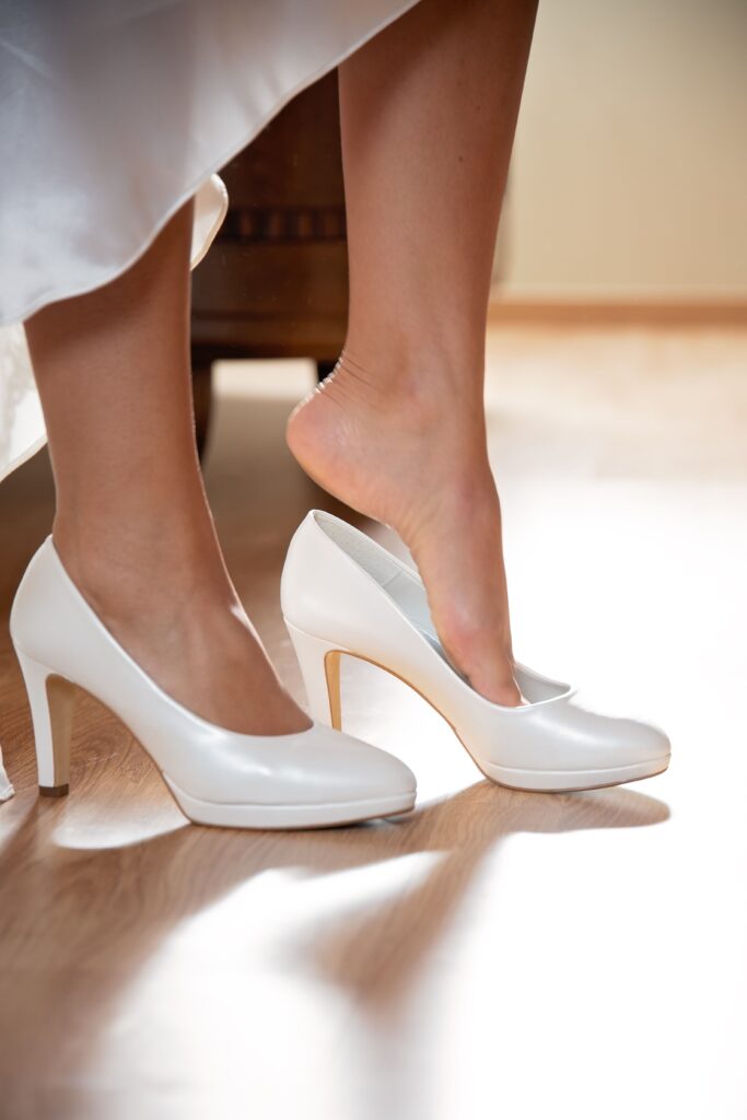
Shoes that are too soft, worn flat, or lack a heel counter can cause the foot to collapse inwards, increasing tension on the medial plantar fascia. Thin-soled sandals, floppy sneakers, and overly flexible flats are common culprits. High heels modify gait and elevate forefoot loading, which can lead to heel spurs and pain in the heel. Extended wear of such shoes heightens the risk of developing conditions like plantar fasciitis. If a shoe allows the heel to move more than a couple of millimeters, it is insufficiently structured.
Recommend Rotating Shoes And Replacing Worn-Out Pairs To Maintain Optimal Foot Care
To prevent heel spurs, it’s essential to switch between at least two pairs of shoes, allowing midsole materials to bounce back and diversifying mechanical strain. Shoes for prevention should include replacing worn-out running shoes every 600 to 1,000 kilometers based on construction and wearer weight. Daily wear shoes must be replaced when heel counters collapse or tread wears unevenly, as these factors can contribute to pain in the heel.
Create A Checklist Of Footwear Features To Look For When Selecting Shoes To Prevent Heel Pain
Checklist: Ensure a correct arch match, a firm heel counter, and adequate cushioning to support the sole. Consider shock-absorbing midsoles and room for toes without slipping. For those suffering from heel spurs, rocker soles can help offload the plantar fascia and reduce pain associated with spurs.
Why Inactive Rest Fails
Extended inactivity feels like it’s safe, but in reality, it tends to exacerbate heel issues by permitting stiffness and weakness to take hold instead of repairing underlying causes.
Inactive rest misses the root cause of your pain and inflammation, so your condition can linger or worsen. When the plantar fascia is left unstretched or unexercised, it shortens and stiffens, continuing to tug on its attachment at the heel bone. That stiffness manifests as pain that frequently subsides after a few minutes of walking, as the tissue warms and lengthens. The pain then returns when you cease and permit the fascia to contract once more. Morning pain follows the same pattern. The fascia shortens overnight, then suddenly stretches on first steps, producing sharp discomfort that rest alone will not stop.
Rest does not activate the body’s repair mechanisms. Mechanical loading in safe ranges stimulates collagen alignment and tissue remodeling. Without that stimulus, healing is slower. Clinical reports demonstrate conservative care can take months. Inactive rest can prolong recovery, sometimes up to 10 months, because muscles and connective tissue remain deconditioned.
Muscle weakness and tightness exacerbate the issue. Calf and Achilles tightness can develop with inactivity and put more strain on the plantar fascia. Tight calves limit ankle dorsiflexion and shift load to the plantar tissues while you walk. Over time, this changes biomechanics, rendering the fascia susceptible to repetitive microtears. Rest does not address these biomechanical flaws, but it merely stops the activity and does not alter the stressing forces.
Active recovery matters: gentle stretching for the plantar fascia and calf, progressive loading exercises, and gradual return to normal activity help remodel tissue and restore function. Targeted therapies like night splints, eccentric calf work, and manual mobilization can be combined with support shoes or orthotics to decrease peak loads. A good shoe spreads the load, pads your heel, and, in combination with exercise, can keep you symptom-free.
Pair rest with focused care and good shoes to compress healing and prevent chronic pain.
Conclusion
You can differentiate the probable cause of heel pain by the pattern of pain, location on the heel, and daily volume of load on your foot. Plantar fasciitis exhibits sharp first-step pain and inner heel pain. Heel spurs demonstrate a pointed bone build-up on X-ray but frequently fail to correlate with pain. Nerve pain sends burning, numb, or electric shocks down the foot. Gait faults, tight calves, and weak hips increase the stress on the heel and keep pain rumbling.
Early, concentrated action produces hard gains. With a guided exam, a few targeted tests, and a clear plan, we sliced his recovery time down significantly. Support your symptoms with night stretch, calf work, paced loading, and shoes with a firm arch. See a clinician if pain persists over six weeks or gets worse. Test one change at a time and keep tabs on what benefits.
Frequently Asked Questions
1. What Is The Most Common Cause Of Heel Pain?
The most common condition is plantar fasciitis, which is inflammation of the band on the sole. It causes sharp first-step pain and is associated with heel spurs and bad foot mechanics.
2. How Can I Tell The Difference Between Plantar Fasciitis And A Heel Spur?
Heel spurs, formed by calcium deposits on the heel bone, are often associated with plantar fasciitis, which causes sharp morning pain and tenderness under the heel.
3. When Should I See A Healthcare Professional For Heel Pain?
See a clinician if pain in the heel persists beyond four to six weeks, limits daily activities, or follows a sudden injury, as early evaluation improves treatment options.
4. Do Orthotics Or Shoe Changes Help Heel Pain?
Yes. For many, custom or supportive over-the-counter orthotics and shoes with good arch support and cushioning can help treat heel spurs and reduce strain.
5. Why Doesn’t Complete Rest Fix My Heel Pain?
Complete rest often does not work due to the underlying biomechanical issues, such as heel spurs. Targeted exercises, including calf muscle stretching, gait correction, and progressive loading, address root causes and prevent recurrence.
6. Can Physical Therapy Or Exercises Treat Heel Pain?
Yes. Stretching, strengthening, and progressive loading programs alleviate pain in the heel and normalize biomechanics, especially for those suffering from heel spurs.
7. Are Imaging Tests Necessary To Diagnose Heel Pain?
Not necessarily. As a rule of thumb, a clinical exam will diagnose plantar fasciitis, which can be related to heel spurs. Imaging such as X-ray, ultrasound, or MRI is used when symptoms are atypical, severe, or not improving with treatment options.
Is Overpronation Affecting The Way You Walk?
Overpronation is more than just a foot issue. When your feet roll inward too much with each step, it can affect your entire body. This imbalance places added stress on your ankles, knees, hips, and lower back, often leading to foot pain, fatigue, poor posture, and recurring discomfort that may seem unrelated to your feet.
At The Shoe Doctor, we identify the root cause of overpronation through advanced 3D foot-mapping technology and detailed gait analysis. By evaluating how your feet move and how your body responds during every step, we gain a clear understanding of your biomechanics. This allows us to pinpoint abnormal movement patterns and determine the best solution to improve your gait and overall alignment.
Once we’ve completed your assessment, we design custom orthotics that provide the right level of support for your unique foot structure and walking pattern. Proper gait correction helps distribute pressure more evenly, improves stability, reduces stress on your joints, and encourages more efficient movement throughout your daily activities.
With more than 20 years of experience, Russell has helped countless people walk with greater comfort, confidence, and stability. If you’re experiencing foot pain, recurring injuries, or discomfort that may be linked to overpronation, schedule your free consultation with The Shoe Doctor today and discover how personalized gait correction can help you move better every day.
Disclaimer
The materials available on this website are for informational and entertainment purposes only and are not intended to provide medical advice. You should contact your doctor for advice on any particular issue. You should not act or refrain from acting based on any content included in this site without seeking medical or other professional advice. The information presented on this website may not reflect the most current medical developments. No action should be taken in reliance on the information contained on this website, and we disclaim all liability for actions taken or not taken based on any or all of the contents of this site to the fullest extent permitted by law.